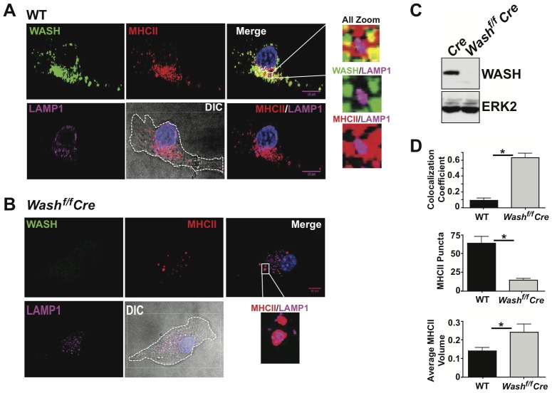
Figure 2. WASH localizes with MHCII in DCs.
BMDCs from (A) Cre and (B) WASHf/f Cre were fixed and labeled with anti-MHCII antibody and WASH for microscopic analysis. (C) Protein lysates prepared from Cre and WASHf/f Cre BMDCs were immunoblotted for WASH and ERK2 (as a loading control). (D) Images from A and B were analyzed for MHCII co-localization with LAMP1 using Pearson's co-localization coefficient. MHCII puncta number and size were quantified by Image J. Zoomed images are demarcated by the white box and lines toward the adjacent image. Differential interference contrast (DIC) images were used to demarcate the outline of the cell. For each condition, >20 individual cells were imaged. Images were collected with 100× oil objective. Scale bars, 10 µm.

