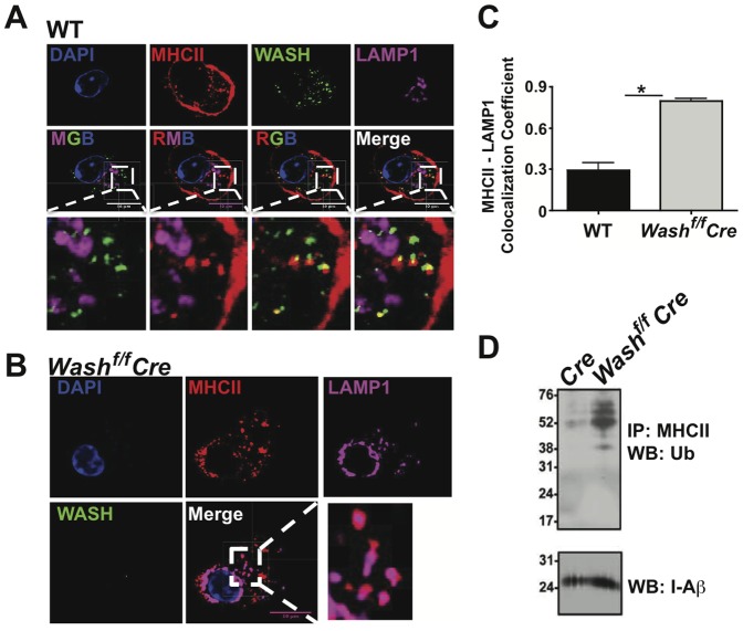
Figure 3. WASH prevents the localization of MHCII into lysosomes following endocytosis.
BMDCs derived from (A) Vav-Cre and (B) WASHf/f Vav-Cre were cultured with an antibody against MHCII following the endocytosis assay then fixed and labeled with antibodies against WASH and LAMP1 for microscopic analysis. (C) Images from A and B were analyzed for MHCII co-localization with LAMP1 using Pearson's co-localization coefficient in ZEN (Carl Zeiss). Zoomed images are demarcated by the white box and dashed lines in the adjacent images. For each condition, >20 individual cells were imaged. Images were collected with 100× oil objective. Scale bars, 10 µm. Bars represent mean ≥ SEM. Horizontal lines indicate statistical comparison between indicated groups, *p≤0.05. (D) Ubiquitinated MHCII was detected in BMDCs by immunoprecipitation of total MHCII followed by immunoblot for ubiquitin (Ub). Blots were subsequently stripped and reprobed with I-Ab antibody as a loading control.

