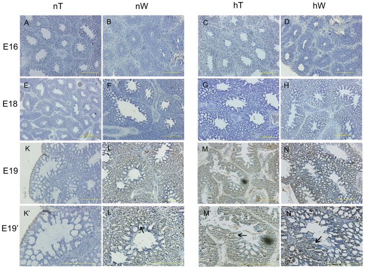
Figure 5. Analysis of apoptosis in lung specimens from chickens incubated under normal and hypoxic conditions.
From E16 to E18, no TUNEL staining was identified (A–H). At E19, apoptotic cells were localized in the mesenchyme surrounding the atrias and infundibula of the chicken lungs (L, M, N). There was no obvious staining in nT chicken at E19 (K). At higher magnification, staining was clearly seen in the regions between ACs and not in the parabronchi, atrias, or infundibula (arrows in L’, M’, N’). hW staining at E19 (arrow in N’) was clearly darker than that observed in hT or nW lung sections at this stage (arrows in M’, N’). The tube density in hW at E19 was also higher than that in sections from those other two groups. Scale bar = 200 µm.

