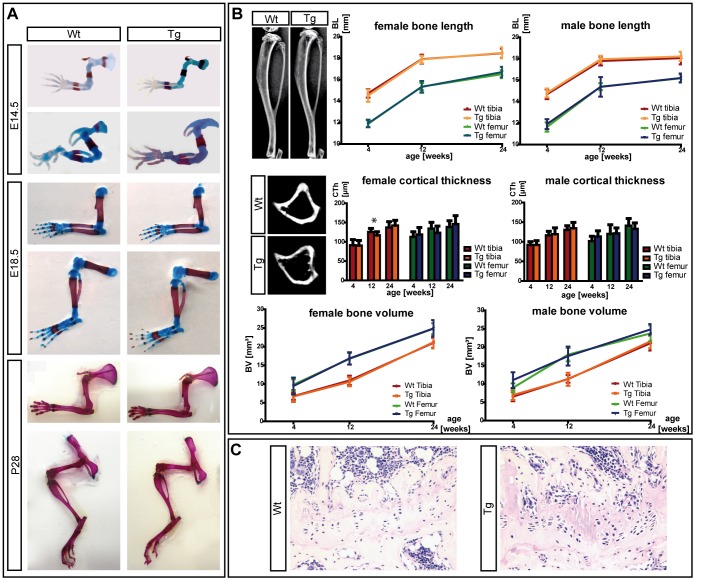
Figure 2. Analysis of postnatal bone parameters of Col2a1-SHOX-transgenic mice.
(A): Alcian Blue/Alizarin Red S staining at different developmental (E14.5, E18.5) and postnatal (P28) stages does not reveal apparent differences between transgenic and wildtype skeletal elements. (B): Postnatal in vivo time-course analysis of bone growth in 65 animals of two transgenic lines by μ-CT analysis. Tibiae and femora of wildtype and Tg(Col2a1-SHOX) littermates at the age of 4, 12 and 24 weeks were scanned, female and male individuals were evaluated separately. Total bone length, cortical bone thickness and bone volume do not show significant differences between wildtype and transgenic females or males. Some transgenic animals presented longer bones and weaker structures of the cortical bone in the subcartilaginous region (indicated in the μ-CT images). Other micromorphological parameters (bone mineral density (BMD), trabecular volume and thickness) showed no significant differences. Statistical analyses were performed using student's t-test. (C): hematoxilin and eosin (H&E) stainings of the growth plate in wildtype and transgenic tibiae. Consistent differences between wildtype and Tg(Col2a1-SHOX) adult growth plates (24 weeks of age) did not exist (N = 8), but some transgenic tibiae showed a buckling, and the columns of chondrocytes became shorter and were not strictly oriented in a parallel assembly compared to the wildtype (right image).

