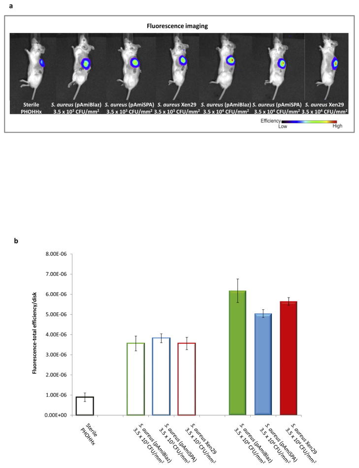
Figure 4.
NIRF imaging approach to detect PHOHHx implant-associated infection with different S. aureus strains at 7 days post-implantation. (a) Bioimaging data of animals scanned in an IVIS® imaging system for in vivo ROS imaging of inflammation associated with implant infection using H-ICG sensor. (b) Quantification of ROS fluorescence data from mice with PHOHHx implants incubated with 3.5 × 10³ CFU/mm2 (open bars), 3.5 × 10⁴ CFU/mm2 (closed bars) and sterile PHOHHx implant (open black bar) (n ≥ 3 mice/time point).

