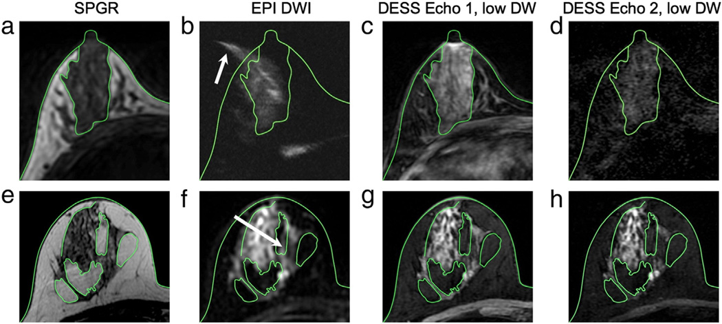
Fig. 6.
Distortion comparison. The green contours show the skin and the interfaces between fat and glandular tissue on the SPGR images (a, e) superimposed on the remaining images. Severe distortion in the spin-echo EPI image (b) can cause some of the signal from the glandular tissue to appear outside of the breast (arrow) when compared to the anatomic reference image (a), but the structures in the DESS images (c, d) correspond well to those in the reference image (a). Even for a high-quality spin-echo EPI image (f), there is still some distortion, and signal from the glandular tissue appears in a fat region (arrow); however, there is no distortion in the DESS images (g, h) when compared to the reference image (e).

