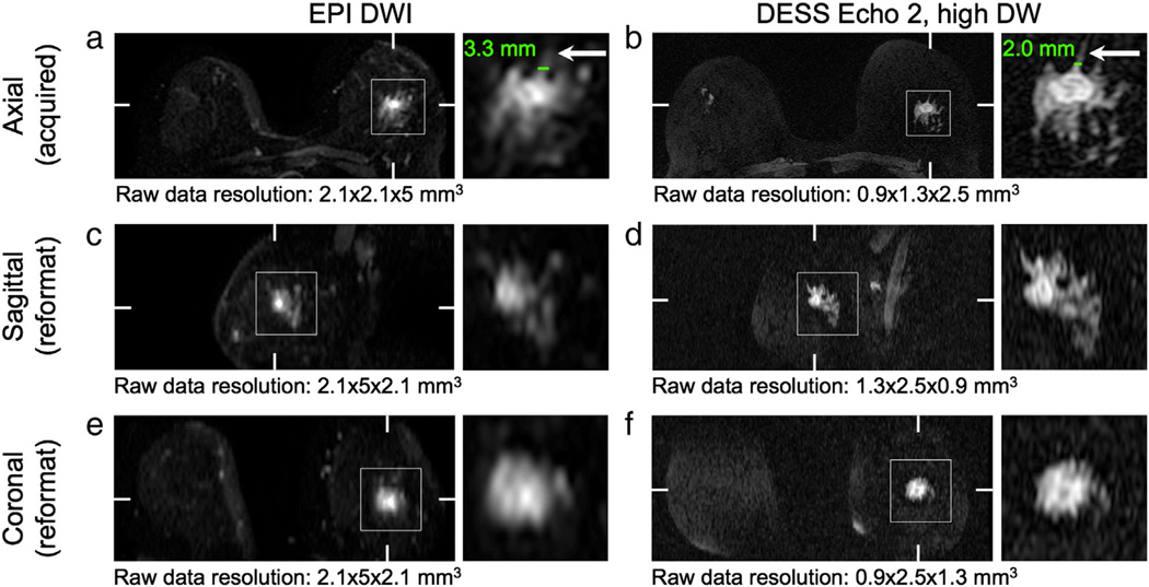
Fig. 7.
Reformatted images. The images acquired with the DESS sequence (b, d, f) have higher resolution than those acquired with the EPI DWI sequence (a, c, e), which is particularly evident in the images reformatted in the sagittal plane (c, d). The higher resolution allows for better depiction of fine features (arrows). Hash marks indicate the locations of the reformatted planes. Detail shows a grade 2 invasive ducal carcinoma.

