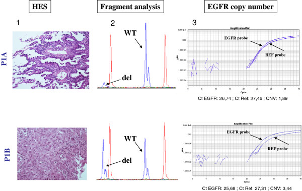
Figure 1.

Illustration of the different patterns of EGFR DEL19 mutation, EGFR CN analyses and the associated histological features for patient P1. -1- HES staining at x100 magnification, P1A : lepidic pattern, P1B: solid pattern. -2- Fragment analysis for EGFR DEL19 mutation. -3- EGFR copy number evaluation. In sample A with lepidic pattern, mutation was validated by a cast PCR assay and there is no increased copy number of EGFR gene (CNV:1.89). In sample B with solid pattern, EGFR amplification was identified (CNV: 3.44).
