Fig. 4.

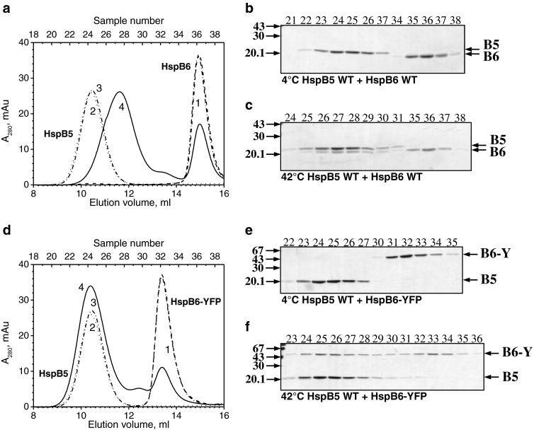
Formation of heterooligomeric complexes of the wild-type HspB5 and either the wild-type HspB6 (a–c) or HspB6-EYFP (d–f). a, d Elution profiles of isolated HspB6 or its fluorescent chimera (1, dashed line), isolated wild-type HspB5 (2, dotted line), and the mixture of two proteins preincubated either at 4 °C (3, dash-dotted line) or 42 °C (4, solid line). b, c, e, f SDS gel electrophoresis of fractions (indicated above each track) collected in the course of SEC. The temperature of preincubation is indicated below each panel. Positions of the wild-type HspB6 (B6), HspB6-EYFP (B6-Y), and the wild-type HspB5 (B5) and molecular weight standards (in kilodaltons) are marked by arrows
