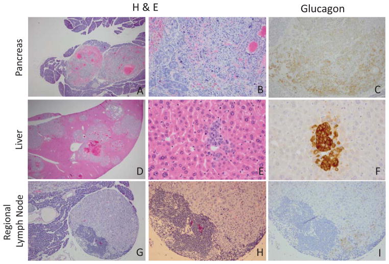
Figure 1.
Hematoxylin and eosin staining of paraffin embedded tissue identifying pancreas tumor and liver and lymph node metastasis in the RenCre/p53/Rb tumor model. A–B. Primary pancreatic tumor (A. 40x, B. 200x) C. Glucagon staining of primary pancreas tumor (200x). DE. H&E metastatic liver (D. 20x, E. 400x). F. Serial glucagon staining of liver metastasis (400x). G–H. Regional lymph node metastasis (G.100x, H.200x). I. Serial glucagon staining of regional lymph node (200x).

