
Figure 1.
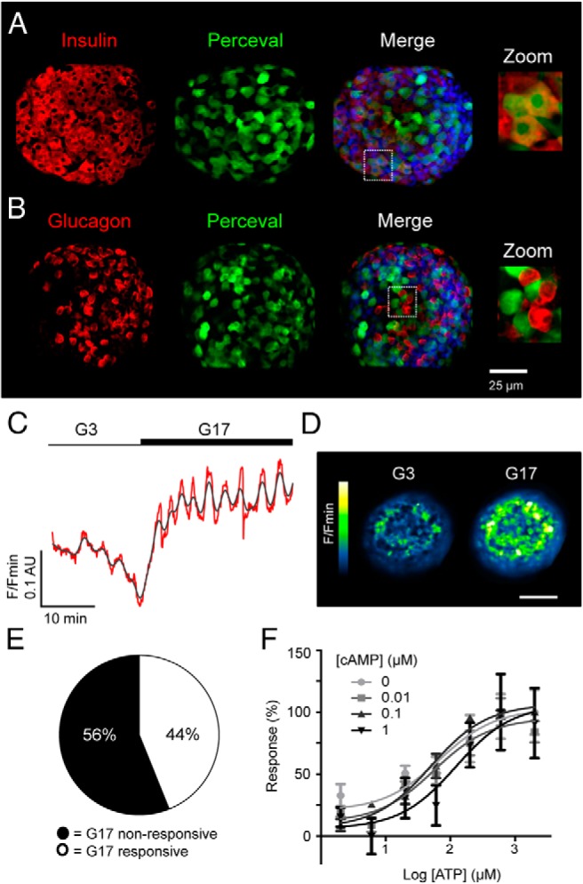
The ATP/ADP-sensor Perceval reports beta cell metabolism in intact islets. A, Specific immunohistochemistry for insulin demonstrates the preferential expression of Perceval in beta cells (DAPI, blue; scale bar shown). B, As for A, but immunostaining against glucagon showing absence of the probe in alpha cells. C, Elevated glucose concentration increases Perceval fluorescence reflecting increases in cytoplasmic ATP/ADP ratio ([ATP/ADP]cyto). D, Representative recording of a Perceval-expressing islet before and during exposure to high glucose (G17; 17 mM glucose) (n = 14 recordings from six animals) (red, raw; gray, smoothed) (scale bar, 50 μm). E, Glucose engages a subpopulation (44%) of metabolically-active beta cells, which respond with [ATP/ADP]cyto increases. F, Dose-response graph depicting ATP-induced increases in dialyzed Perceval fluorescence, which are unaffected by the presence of increasing cAMP concentrations (0–1 μM) (sigmoidal dose-response fitted to 2–3 independent repeats).