
Fig. 3.
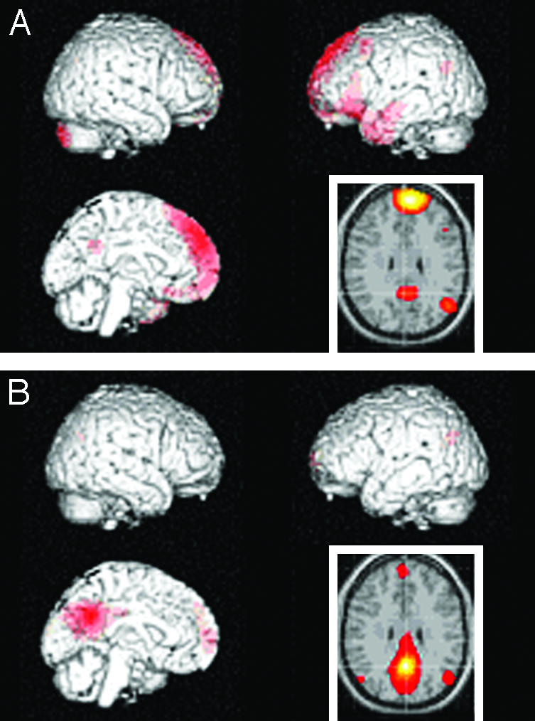
Connectivity patterns during combined tasks. The data are shown as z-score maps of synchronous activity in the rest of the brain across conditions with medial prefrontal region (A), and with medial parietal/posterior cingulate region (B). The former is mainly connected with medial parietal/posterior cingulate, left temporoprefrontal region, and left inferior parietal region; the latter is mainly connected with medial prefrontal cortex and bilateral inferior parietal cortices.