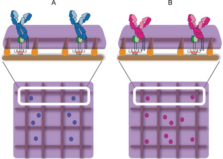
Figure 1.
Schematic presentation of IgM-BCRs and IgG-BCRs. (A) IgM-BCRs composed of membrane-bound forms of IgM (light blue indicates the light chain, dark blue indicates the heavy chain) associated with a heterodimer of Igα and Igβ (green) on a mature naïve B cell. The mIg and the Igα/Igβ heterodimer are associated through non-covalent interactions. (B) IgG-BCRs composed of membrane-bound forms of IgG (light pink indicates the light chain, dark pink indicates the heavy chain) associated with the Igα/Igβ heterodimer (green) on a memory B cell. In A and B, the BCRs are depicted in compartmentalized plasma membrane regions that are maintained by membrane-proximal actin fence and anchored transmembrane protein pickets as proposed by Kusumi et al.79,80,81. The ITAM-containing cytoplasmic tails of Igα/Igβ heterodimer within the IgM- or IgG-BCR complexes are also shown.

