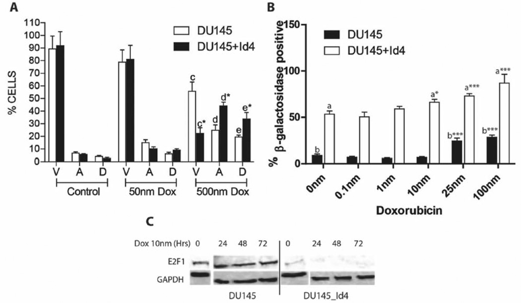
Figure 3.
Effect of doxorubicin on apoptosis and senescence in DU145 and DU145+Id4 prostate cancer cell lines. A: Flow cytometric data show the effect of 0 (control), 50 and 500 nm doxorubicin treatment (24 h) on apoptosis in DU145 and DU145+Id4 cells after AnnexinV – Alexa488 and PI staining. The viable (V), apoptotic (A) and dead (D) cells are represented (mean±SEM of three experiments in duplicate). Apoptosis in DU145 and DU145+Id4 cells treated with 50 nm doxorubicin was not statistically different, whereas a significant increase in apoptosis was observed in both cells types with 500 nm doxorubicin. *p<0.001, Student’s t-test. B. DU145 and DU145+Id4 cells were treated with 0–100 nm doxorubicin and stained with SA-b-galactosidase. The blue nuclei due to SA-b-galactosidase staining were counted in 15 randomly-selected fields and are expressed as mean±SEM. The blue SA-b-galactosidase-positive nuclei were counted and expressed as mean+SEM. ***p<0.001, t-test for columns “a” and “b”. C: Western blot analysis of E2F1 expression in DU145 and DU145+Id4 cells treated with 10 nm doxorubicin for 0, 24, 48 and 72 h.

