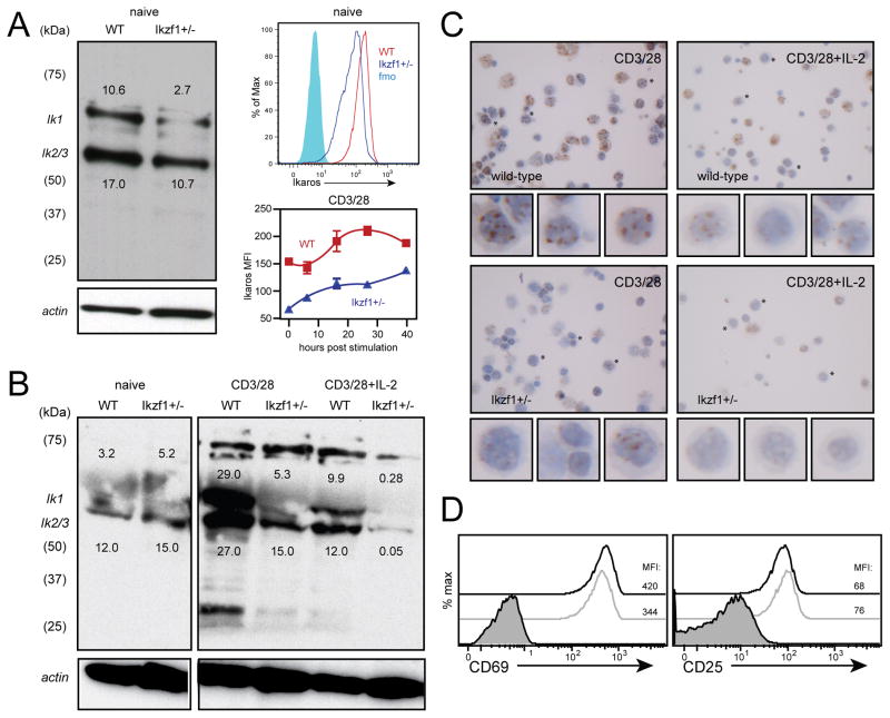
Fig. 2. CD8+ T cells hemizygous for the Ikzf1 gene exhibit reduced Ikaros protein expression.
Polyclonal naive CD8+ T cells sorted by CD62L+CD44- from wild-type or Ikzf1+/− mice (1×10⁶ cell equivalents) were subjected to immunoblot analysis of Ikaros protein expression as in Fig. 1 (A, left panel). Ikaros expression by naive RAG1−/− OT-1 (WT, red) or RAG1−/− OT-1 Ikzf1+/− CD8+ T cells (blue) was measured by flow cytometry (A, right panels, gated on CD62L+CD44-CD8+), immunoblot analysis (B), or immunohistochemistry (C) following stimulation with plate-bound anti-CD3/CD28 in the presence or absence of IL-2 (10 ng/ml). D. CD69 and CD25 expression by 12 hour anti-CD3/CD28-stimulated RAG1−/− OT-1 (black) or RAG1−/− OT-1 Ikzf1+/− (grey) CD8+ T cells was measured by flow cytometry (gated on live CD8+ T cells). Numbers above the Ik1 band and below the Ik2/3 band indicate pixel density (x1000). Data are representative of two independent experiments.

