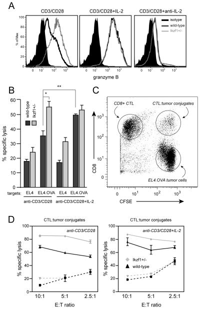
Fig. 6. Loss of Ikaros function leads to enhanced cytolytic capacity by CD8+ T cells.
A. Naïve-enriched RAG1−/− OT-I (dark gray) or Ikzf1+/− OT-I (light gray) CD8+ T cells were stimulated with plate-bound anti-CD3 and anti-CD28 for 48 hours in the presence or absence of IL-2 or anti-IL-2. PMA (30 ng/ml) and ionomycin (1uM) were added for the last four hours of culture. Expression of granzyme B was assessed by flow cytometry. Data are representative of two independent experiments. Filled black histograms - granzyme B FMO negative control. OT-I cells (dark gray) or Ikzf1+/− OT-I cells (light gray) were stimulated for 48 hours as in A, rested in medium overnight, then mixed at a 10:1, 5:1 and 2.5:1 ratio with CFSE-labeled EL4 or EL4.OVA targets for 3 hours. Viability of bulk CFSE+ EL4 or EL4.OVA targets at a 10:1 E:T ratio is shown in (B) or in conjugates with CD8+ T cells (CFSE+CD8+ gate in C) at all ratios in (D). Dashed lines indicate response to EL4. Data are representative of 3 independent experiments. Statistical significance was determined by Student’s T-test for triplicate values/group in B - * p<0.05, ** p<0.001, *** p<0.0001. Values plotted in D represent mean+/−SEM of duplicate cultures.

