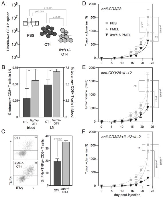
Figure 7. Enhanced in vivo effector function by Ikzf1+/− CD8+ T cells.
Naive RAG1−/− OT-1 (dark grey) and Ikzf1+/− RAG1−/− OT-1 (light grey) CD8+ T cells were purified, primed with plate-bound anti-CD3/28 Ab for 48 hours, and 5×10⁶ cells were adoptively transferred into B6 mice. Two hours later, mice were challenged with 1×10⁵ LM-OVA, and blood, LN and spleens were harvested at day 3 post-infection. Live LM-OVA in the spleen (A) and MHC-OVA tetramer+ CD8+ T cells in blood and LN (B) were enumerated. LN cells were restimulated ex vivo with OVA peptide (1 μM) and analyzed by flow cytometry for production of IFN-γ and TNFα (C). Recipients: PBS, n=3; RAG1−/− OT-1, n=4; Ikzf1+/− RAG1−/− OT-1, n=3. Statistical significance was determined by Student’s T-test (A) and one-way ANOVA (B, C) and p values are indicated. For tumor studies, naive PMEL (grey) or Ikzf1+/− PMEL (black) CD8+ T cells were purified, stimulated in vitro with plate-bound anti-CD3/28 Ab (D), or with the addition of IL-12 (E) or IL-2 and IL-12 (F), and 1×10⁶ cells were adoptively transferred into B6 mice. Mice were challenged subcutaneously 24 hours later with 1×10⁵ B16 melanoma cells. Tumors: PBS, n=6; PMEL, n=6; Ikzf1+/− PMEL, n=6. Statistical significance was determined by two-way ANOVA and p values are indicated. * p<0.05, ** p<0.001, *** p<0.0001.

