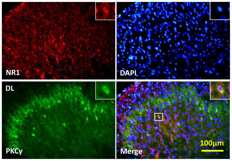
Fig. 5. Co-localization of spinal NR1 and PKCγ expression.
There was co-localization of NR1 and PKCγ immunoreactivity in the superficial layers (I & II) of the spinal cord dorsal horns at the lumbar (L4) level. Spinal cord samples were taken from burn-injured young rats receiving a combination of morphine and midazolam treatment for 14 days (n=3). Blue: DAPI for nucleus. Scale bar: 100 μm. DL: the dorsolateral part of the spinal cord dorsal horn.

