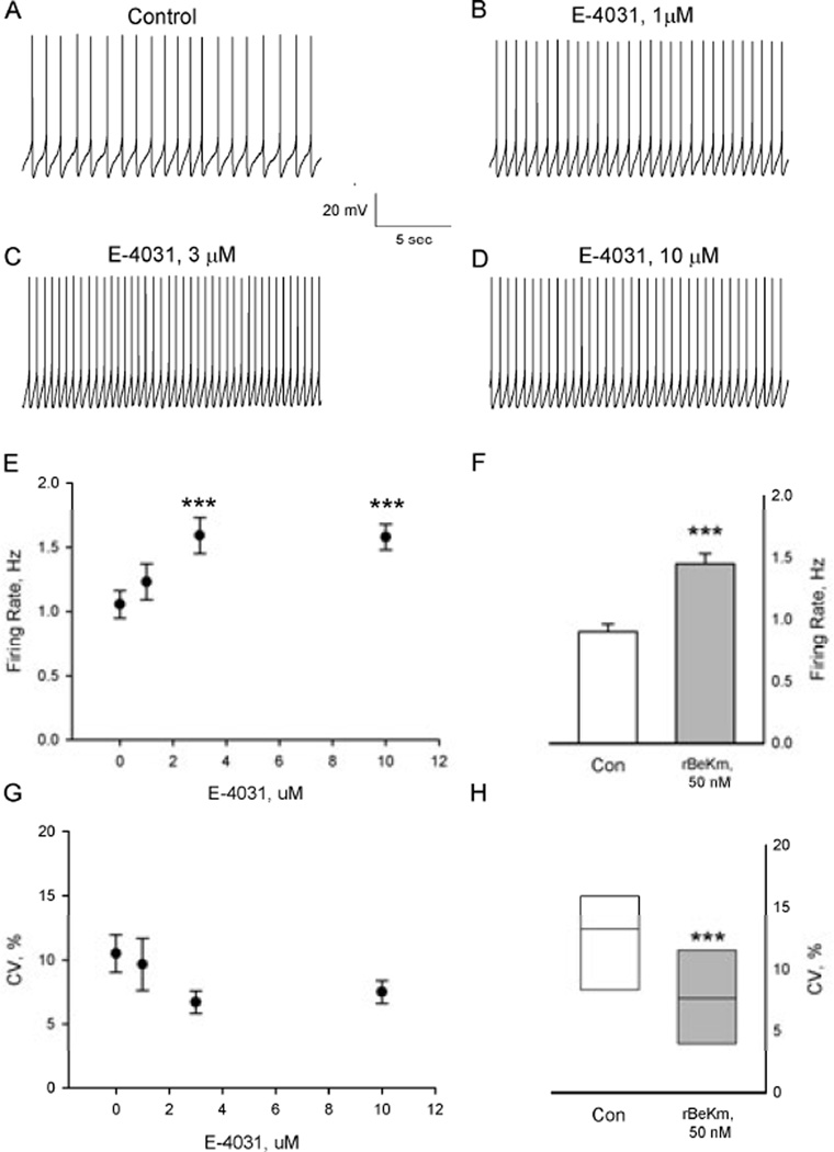
Figure 1.
Blockade of ERG K+ channels increased the firing rate of DA-containing neurons in brain slices. (A–D) Representative tracings from a spontaneously active DA neuron treated with increasing concentrations of E-4031 (1–10 µM). All records are from the same cell. (E) Summary of the effects of cumulative administration of E-4031 on DA cell firing rate. Each point represents the mean ± SEM of n=8 neurons. *P < 0.001 vs. control (Bonferroni t test). (F) Bath application of rBeKm-1 (50 nM) significantly increased the activity of DA neurons (***P < 0.001, paired t-test, n=8). (G and H) Effects of (G) E-4031 and (H) rBeKm-1 on the interspike interval coefficient of variation. The horizontal line within the box plots indicates the median value while the upper and lower limits of the box denote the 75th and 25th quartiles, respectively. ***P < 0.05, Wilcoxon signed-rank test.

