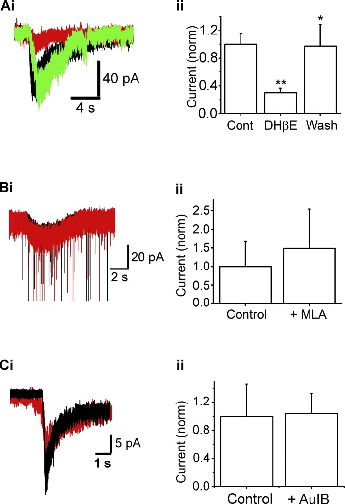
Fig. 4.
Functional α4β2-nAChRs are expressed on ET cells. Ai: raw trace of ACh/At-induced current in an ET cell under Cont conditions (black), in the presence of 10 μM dihydro-β-erythroidine hydrobromide (DHβE; red), and after washout of DHβE (green). Cell was held at −70 mV. Aii: summary of effect of 10 μM DHβE on ACh/At-induced currents normalized to currents under Cont conditions (**P < 0.01, *P < 0.05). n = 9 (Cont and DHβE); n = 4 (Wash). Bi: 1-s, 1 mM ACh/At (starts at arrow) results in a slow inward current under Cont (black trace) as well as in the presence of 10 nM methyllycaconitine (MLA; red), at −70 mV. Some sEPSCs have been truncated. Bii: MLA application does not significantly alter the ACh/At-induced current (n = 5, P > 0.2). Ci: nAChR current response in the absence (black) and presence (red) of 10 μM CTx-AuIB. The current was not significantly inhibited. Cii: mean values from 6 cells. The presence of CTx AuIB did not significantly attenuate ACh/At responses, suggesting the α3β4*-nAChR subtype is not a major contributor to the nAChR currents on ET cells.

