Fig. 1.
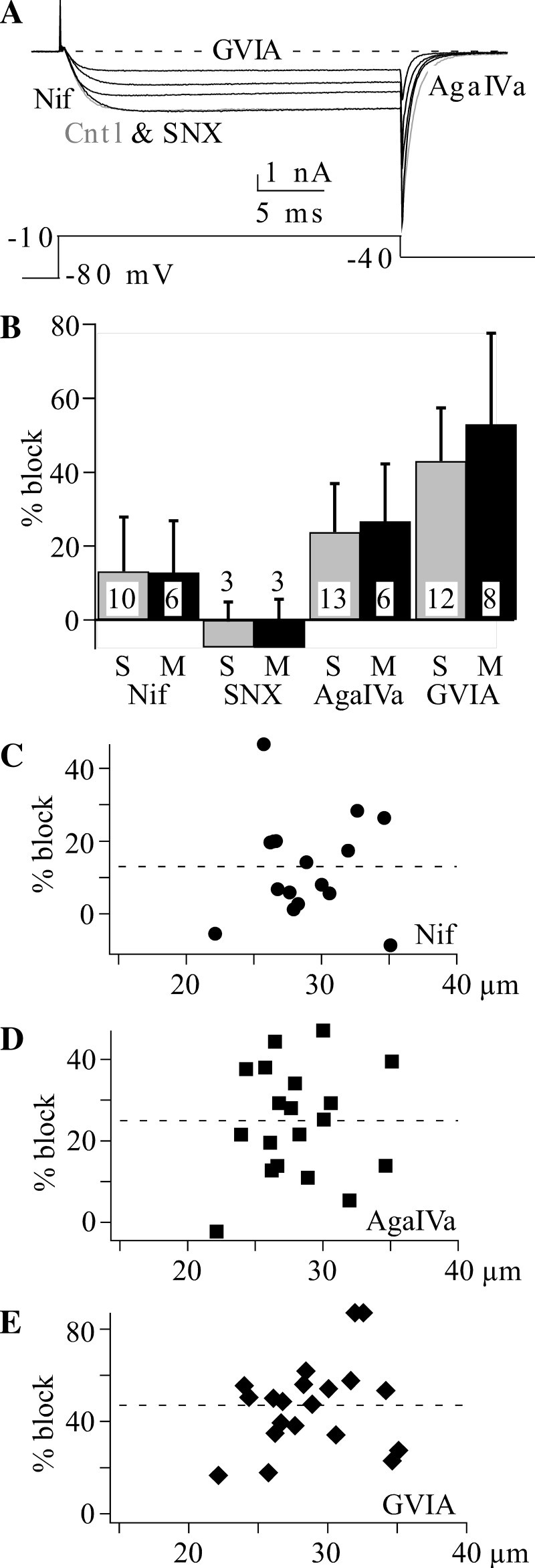
Voltage-dependent calcium (CaV) 2.2 channels generate the dominant CaV current in muscle afferent neurons. CaV current in 1,1′-dioctadecyl-3,3,3′,3′-tetramethylindocarbocyanine perchlorate (DiI) labeled muscle afferent neurons was measured using 5 mM Ba2+ external solution. A: example currents showing the effect of 0.3 μM SNX482 (SNX), 0.2 μM ω-agatoxin IVa (AgaIVa), 3 μM nifedipine (Nif), and 10 μM ω-conotoxin GVIA (GVIA). The voltage protocol is shown below the current traces. Cntl, control. B: comparison of mean ± SD calcium current block in small (S; 20–30 μm) vs. medium (M; 30–40 μm) muscle afferent neurons by the indicated blockers. The blocker concentrations are the same as indicated for A. C–E: the distribution of percentage block vs. neuron diameter for 3 μM Nif (C), 0.2 μM AgaIVa (D), and 10 μM GVIA (E). Note the different y-axis scale for GVIA. The dashed line indicates average block.

