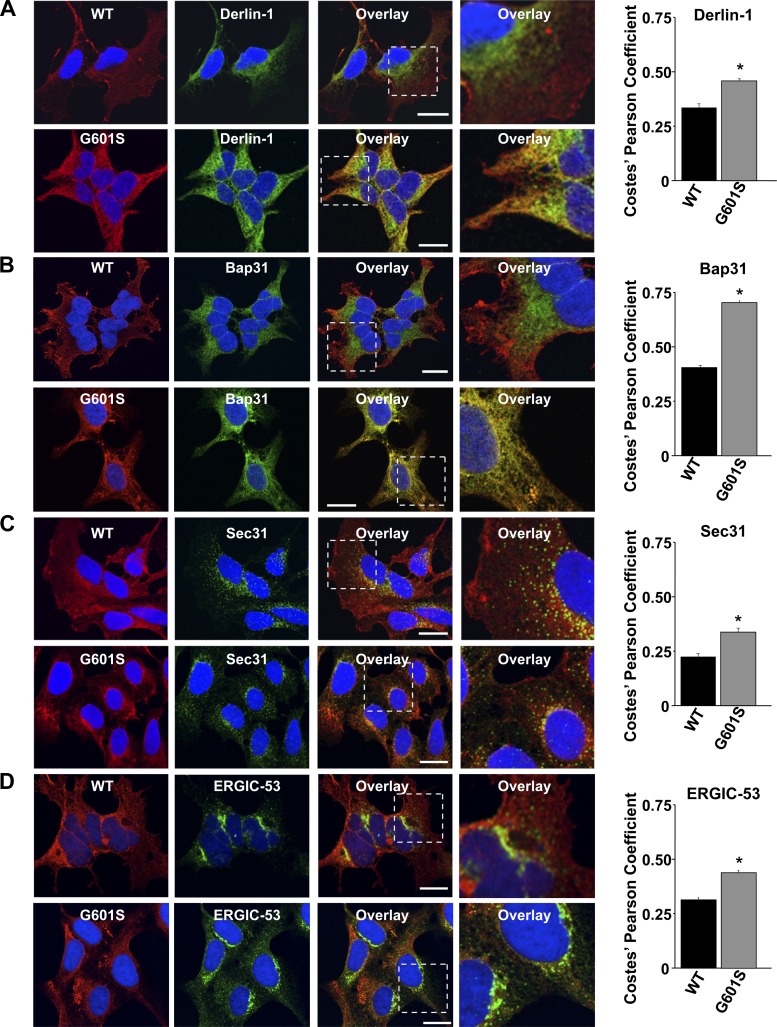
Fig. 1.
G601S selectively colocalizes with Bap31. Shown are representative fluorescent images of cells expressing wild-type (WT)-Kv11.1 or G601S immunostained with anti-Kv11.1 (red, first column) and anti-Derlin-1 (A), anti-Bap31 (B), anti-Sec31 (C), or anti-ERGIC-53 (D) (green, second column). The overlay between anti-Kv11.1 and the different endoplasmic reticulum (ER) markers is shown in the third column. Scale bars, 10 μm. The fourth column shows a portion of the overlays (white dashed box, third column) in greater detail. Also shown are the corresponding Costes' Pearson coefficients (*P < 0.05 compared with WT-Kv11.1).

