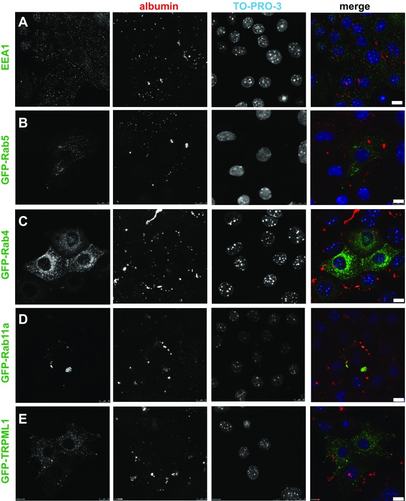
Fig. 3.
Endocytic marker distribution in PTCs. Filter-grown PTCs [transfected with the indicated green fluorescent protein (GFP)-tagged markers where indicated] were incubated with 40 μg/ml Alexa Fluor 555-albumin added apically for 30 min (red), fixed, and then processed for immunofluorescence. Maximal projections of albumin labeling are shown to highlight the endocytically active cells, along with a single optical slice showing distribution of endocytic/lysosomal markers and the nuclear stain TO-PRO-3. Scale bars, 10 μm. EEA1, early endosome antigen 1; TRPML1, transient receptor potential cation channel mucolipin subfamily, member 1.

