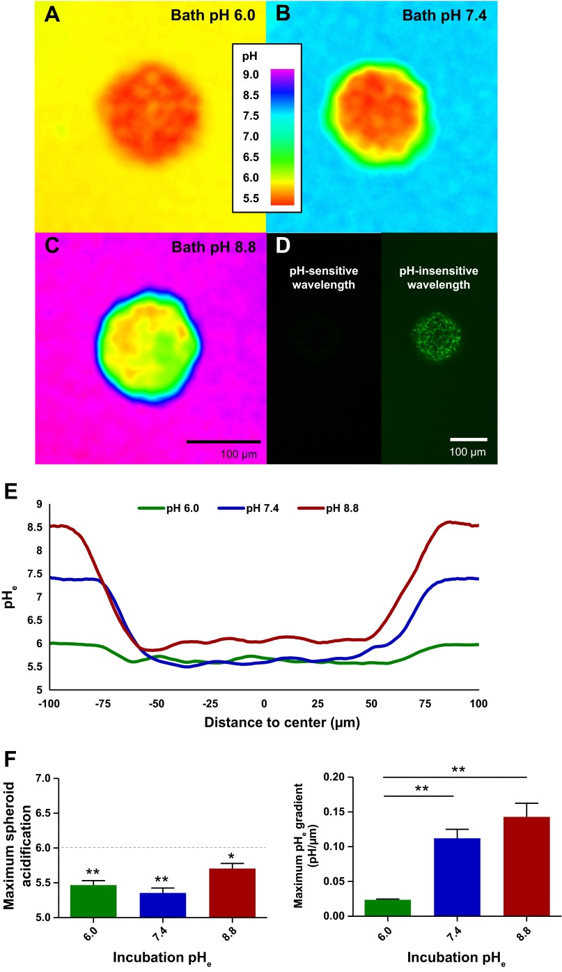
Fig. 1.
Quantification of tumor spheroid acidification. A–C: >1-wk-old U251 human glioma spheroids were preincubated and then placed for confocal imaging in pH 6.0, 7.4, and 8.8 baths with 20 μM cell-impermeant seminaphtharhodafluor-5F 5-(and-6-)-carboxylic acid (SNARF-5F) pH indicator dye. Representative z sections are shown at ×20 magnification through the middle of the spheroids. D: full diffusion of SNARF-5F in a representative pH 6.0-bathed spheroid. E: line graphs through the center of the representative spheroids show that they create standing extracellular pH (pHe) gradients that can acidify to ∼pHe 6.0 regardless of bathing medium. F and G: quantification of maximum acidity (1-tailed t-test for H0 pHe < 6.0, n = 3) and maximum gradient of acidification (1-way ANOVA with Tukey-Kramer post test, n = 3), respectively. *P < 0.05; **P < 0.01.

