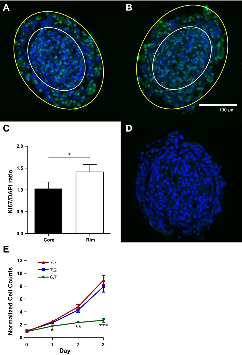
Fig. 2.
Human glioma spheroids evolve pHe and proliferation gradients. A and B: >1-wk-old U251 human glioma spheroids were embedded in paraffin, cross-sectioned, and stained for Ki67 (green) and 4′,6-diamidino-2-phenylindole (DAPI, blue). White outline encapsulates the core of the spheroid, while the area between yellow and white outlines encapsulates the spheroidal rim. Magnification ×20. C: Ki67 staining normalized to DAPI shows more proliferation of the rim glioma cells (2-tailed paired t-test, n = 7). D: control spheroid without Ki67 primary antibody. E: quantification of Coulter counter data, with cells cultured at varying pHe levels and media changed daily (2-tailed t-tests vs. pHe 7.2 as control, n ≥ 6). *P < 0.05; **P < 0.01; ***P < 0.001.

