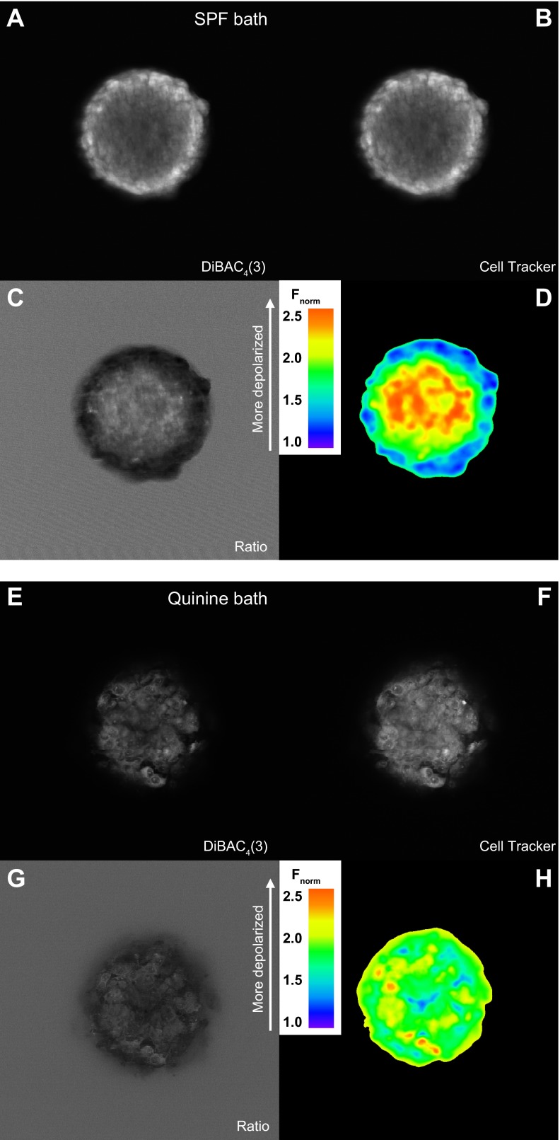
Fig. 6.
Quinine depolarizes glioma spheroids. A–D: confocal cross-section image of a U251 glioma spheroid coloaded with the voltage-sensitive dye bis-(1,3-dibutylbarbituric acid) trimethine oxonol [DiBAC4(3); A] and the cell-tracking dye CellTracker Orange (B). Images were normalized to CellTracker Orange (Fnorm; C) and then falsely colored (D). E–H: confocal imaging of a U251 spheroid preincubated for 2 h with 1 mM quinine. E: voltage-sensitive channel; F: cell-tracker channel; G: normalization to cell-tracking dye; H: ratiometric coloring.

