Figure 4.
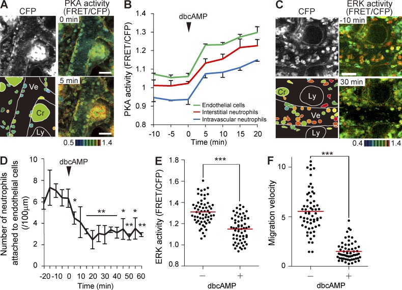
PKA inhibition of ERK activity, recruitment to endothelial cells, and migration of neutrophils. (A) FRET images of the lamina propria of the intestinal mucosa in PKAchu mice pretreated with LPS and fMLP. A cAMP analogue, dbcAMP, was injected intravenously at 0.3 g/kg at time 0. The bottom left panel shows the schematic view of this region. Cr, crypt; Ly, lymphatic vessel; Ve, venule. Gamma, 1.7. The image is representative of a mouse in three independent experiments. (B) Time courses of the PKA activity of intravascular and interstitial neutrophils and endothelial cells. In each of three mice, 10 neutrophils in and out of the venules and 10 endothelial cells were randomly selected in the CFP images and examined for PKA activity. Mean data of three mice are shown with one SD. (C) FRET images of the lamina propria of the intestinal mucosa in Eisuke mice pretreated with LPS and fMLP. A cAMP analogue, dbcAMP, was injected intravenously (0.3 g/kg) at time 0. Images are cropped from Video 5. Gamma, 1.2. The image is representative of a mouse in three independent experiments. (A and C) Bars, 30 µm. (D) Inhibition of the neutrophil attachment to the endothelial cells by dbcAMP treatment. The number of neutrophils on the endothelial cells was counted in three mice, and the mean value and one SD of the average of each mouse are plotted against time. Asterisks indicate the result of the paired Student’s t test between each time point and time 0: *, P < 0.05; **, P < 0.01. (E and F) Correlation of ERK activity and migration velocity of interstitial neutrophils before and after dbcAMP treatment. 60 neutrophils that were imaged in three mice were randomly selected in the CFP images before and after dbcAMP treatment and examined for their ERK activity (E) and migration velocity (F) during 5 min of time-lapse imaging. Red bars indicate the mean values. ***, P < 0.001 (E, Student’s t test; F, Mann–Whitney U test).

