Figure 2.
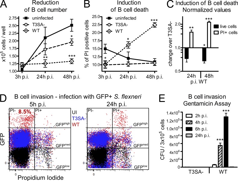
S. flexneri induces B cell death dependent on the T3SA in vitro. The human IgA+ CL-01 B cell line was infected for 30 min with WT or T3SA− bacteria before addition of gentamicin. (A) Count of in vitro–infected human CL-01 B cells over time. Asterisks indicate statistical difference to the uninfected control. (B) Percentages of in vitro–infected PI+ human CL-01 B cells over time. Asterisks indicate statistical difference to the uninfected control. (C) Fold changes of live cell number and percentage of PI+ cells are presented for WT infection over infection with the T3SA− mutant as normalized values. Asterisks indicate statistical difference to the T3SA− strain. (D) Flow cytometry analysis of cells infected with GFP-expressing bacteria. GFPhigh PI− B cells were detected 5 h p.i. with WT, but not T3SA− bacteria (P < 0.001), representing 8.47 ± 1.1% (mean ± SEM) invaded cells. At 24 h p.i., GFPhigh cells are PI+. (E) Invasion assay for CL-01 B cells. The number of CFUs per 3 × 10⁵ infected cells is presented for WT and T3SA− bacteria at 2, 4, 6, and 24 h p.i. Three independent experiments were performed in triplicate for A–E and data are presented as mean ± SEM. Statistically significant differences were determined by two-way ANOVA with Bonferroni post-test. *, P < 0.05; **, P < 0.01; ***, P < 0.001.

