Figure 6.
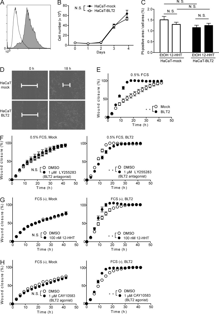
12-HHT and a synthetic BLT2 agonist enhance HaCaT-BLT2 cell migration. (A) Flow cytometry analysis of HaCaT cells stably expressing Flag-tagged BLT2 (gray) and HaCaT-mock transfectants (white) after staining with anti-Flag antibody. (B) Growth of HaCaT-mock and HaCaT-BLT2 cells (n = 3 experimental replicates). (C) Quantification of cell death of HaCaT-mock and HaCaT-BLT2 cells by propidium iodide (PI) staining 12 h after scratching. Cells were cultured in medium containing EtOH or 100 nM 12-HHT (n = 3–4 experimental replicates). (D) Representative fields show the wound gap filled by HaCaT-mock and HaCaT-BLT2 cells cultured in medium containing 0.5% FCS at 0 h and 18 h. (E–H) Quantification of HaCaT-mock and HaCaT-BLT2 cell migration (n = 4 experimental replicates). The cells were cultured in medium containing 0.5% FCS (E and F) or in FCS-free medium (G and H) containing the synthetic BLT2 antagonist LY255283 (F), 12-HHT (G), or the synthetic BLT2 agonist (H). Data represent the mean ± SEM. **, P < 0.01; N.S., not significant (B and E–H, two-way ANOVA; C, two-way ANOVA with Bonferroni post hoc tests). All the results are representative of at least two independent experiments.

