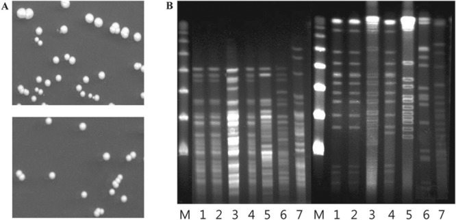
FIG 2.

Colony morphologies and PFGE analysis of M. massiliense isolates. (A) Both sputum (top) and blood (bottom) isolates grew smooth white colonies, 2 mm in diameter, on Middlebrook 7H10 agar after incubation for 7 days at 37°C. (B) PFGE analysis of genomic DNA from bacterial isolates after digestion with XbaI (left set of lanes) and DraI (right set of lanes). The patient's sputum (lanes 1) and blood (lanes 2) isolates show identical profiles, while three M. massiliense isolates (lanes 3, 4, and 5) and two M. abscessus sensu stricto isolates (lanes 6 and 7) from epidemiologically unrelated patients each had distinct profiles. Lanes M, a lambda ladder PFGE marker (New England BioLabs, Ipswich, MA) was used as a size marker.
