Figure 2.
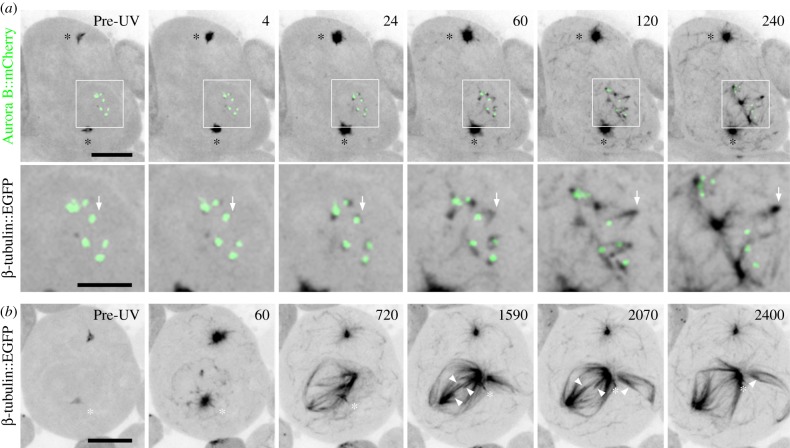
Functional acentrosomal spindles form from MTs nucleated within the nucleus and at kinetochores. (a) Sequence showing MT regrowth in a wild-type spermatocyte expressing both β-tubulin56D::EGFP and Aurora B::mCherry to label MTs and kinetochore position, respectively. Prior to the UV pulse, no MTs are detected in the low magnification or boxed and zoomed panels. After the pulse, MTs emanate from the centrosomes (*) and form throughout the cytoplasm. Within the nucleus MTs appear at the persisting membranes, in the nucleoplasm and directly adjacent to the kinetochores. These latter MTs first appear as foci but rapidly assume a ‘v’ shape as they extend away from the kinetochore led by their vertices (arrows follow formation and extension of a single example). Non-kinetochore nucleated MTs undergo similar movements. Both populations can fuse into larger more intricate structures. (b) Sequence from an increased duration recording of an EGFP-tagged tubulin expressing cell following MT regrowth. In this cell, a functional, multi-polar spindle forms. Although one centrosome is proximal to the nucleus (*), it does not appear to contribute substantial numbers of MTs and instead travels along the acentrosomal half-spindle towards its pole. As the spindle matures (1590), k-fibres are observed (arrowheads) which eventually shorten (2070–2400) similar to those in anaphase controls. Time is in seconds relative to the UV pulse. All images are z-projections. Bars are 10 and 5 μm in low magnification and zoomed panels, respectively.

