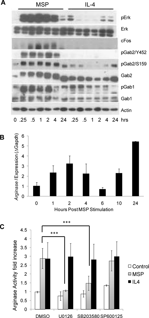
Figure 3. MSP induces arginase I expression in primary macrophages in a Map kinase-dependent manner.
A) Resident peritoneal macrophages were either unstimulated, or stimulated with 100 ng/mL MSP or 10 ng/mL IL-4 for the indicated times. Cell lysates were isolated and levels of phosphorylated Erk (pErk), total Erk, cFos, phosphorylated Gab2 (pGab2Y452 and pGab2S159), total Gab2, phosphorylated Gab1 (pGab1Y307), total Gab1, and actin were assessed by Western blot analysis. Data are representative of two or more independent experiments. B) Resident peritoneal macrophages stimulated with 100 ng/mL MSP for the indicated times and RNA was collected for quantitative RT-PCR analysis. Data are presented as mean ± S.D. and are representative of four independent experiments. C) Resident peritoneal macrophages were stimulated with DMSO, 10 µM U0126, 10 µM SB203580 or 20 µM SP600125 for 2 hours followed by stimulation for 24 hours with 100 ng/ml MSP or 5 ng/ml IL-4 and arginase activity was assessed. Results are the average of two independent experiments. ***p<0.001.

