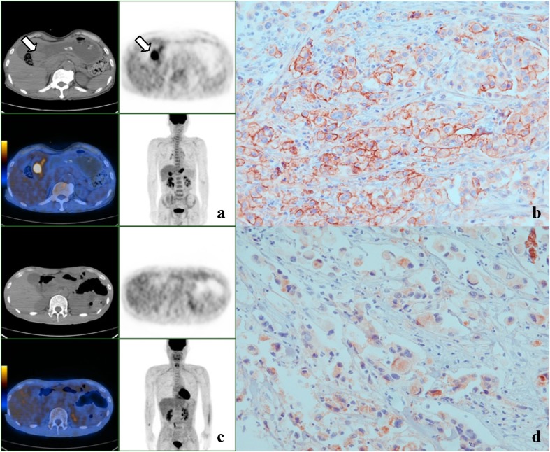
Fig. 1 a–d.
Representative PET and immunohistochemical staining images for GSRC with GLUT-1 expression in membrane and cytoplasm. a, b A case of stage IV GSRC showing intense FDG uptake (SUVmax 8.4) in the gastric antrum (arrow) and membranous GLUT-1 staining. c, d Another case of stage IV GSRC showing no significant FDG uptake in gastric wall and cytoplasmic GLUT-1 staining (×400)

