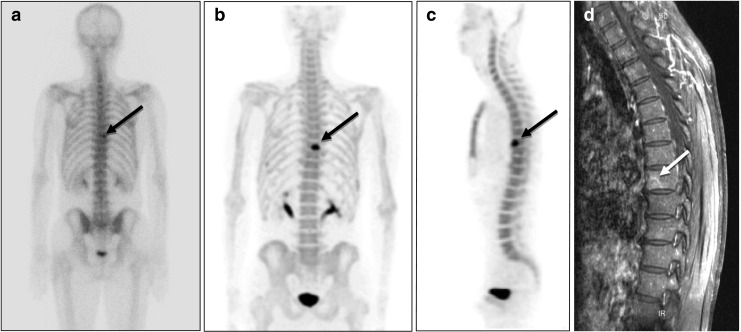
Fig. 1.

18F-fluoride bone PET of a 61-year-old male patient with prostate cancer. a A suspected to have bone metastasis in the T-8 area (black arrow) in a 99mTc-HDP bone scan (posterior whole body image). b18F-fluoride bone PET readily revealed bone metastasis at the T-8 vertebral body in a maximum intensity projection (MIP) posterior view image, and c in a sagittal image. d T1-weighted gadolinium-enhanced MRI also demonstrated metastasis at the T-8 vertebral body (white arrow). Radiation therapy was applied to the lesion, and a follow-up bone scan revealed improvement
