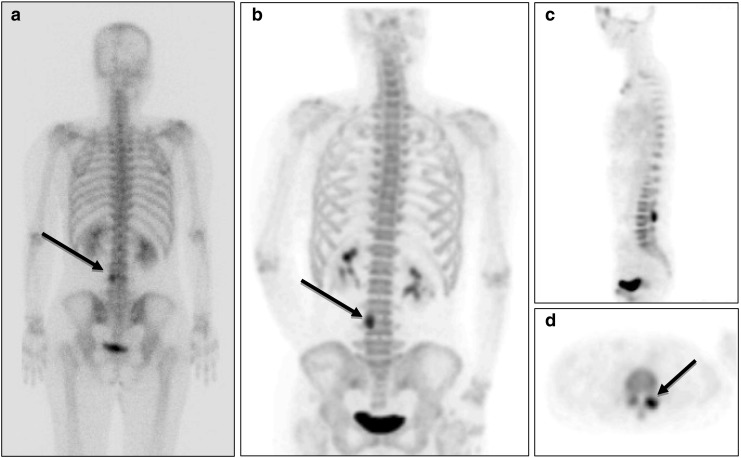
Fig. 2a–d.

18F-fluoride bone PET ruled out the presence of bone metastasis. a A 45-year-old female patient with breast cancer was suspected of having bone metastasis at L-4 (black arrow) in a 99mTc-HDP bone scan (posterior whole body image). b18F-fluoride bone PET indicated that the lesion was located at left L3-4 facet joint in a MIP posterior view image, c a sagittal image, and d a transaxial image. A follow-up bone scan conducted at 17 months after the bone PET study did not show any change in the lesion, and the patient showed no clinical evidence of bone metastasis
