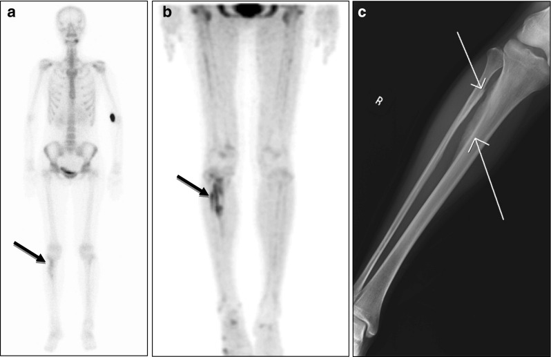
Fig. 5a–c.

18F-fluoride bone PET depicted more intense and more extensive reactive bone lesions than 99mTc-HDP bone scan. a A 25-year-old female patient with intramuscular hemangioma in the right tibialis posterior muscle was found to have reactive bone lesions in the right tibia and fibular by 99mTc-HDP bone scan (black arrow). b However, 18F-fluoride bone PET demonstrated more intensive and extensive bone lesions. c Simple radiography visualized reactive bone lesions of the right tibia and fibular (white arrows)
