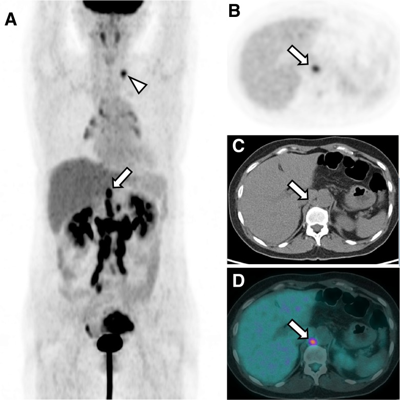
Fig. 2.
Representative PET/CT image of an ovarian cancer patient with RCLN and supra-diaphragmatic LN metastasis MIP image of 18F-FDG PET (a), transaxial 18F-FDG PET image (b), non-contrast-enhanced CT (c), and rigid fusion image (d) of a patient with left ovarian cancer. The images show a left ovarian mass with increased metabolic activity, multiple hypermetabolic LNs at the retroperitoneal area, and a hypermetabolic LN at the left supraclavicular fossa (arrowhead), but no evidence of distant organ involvement. The hypermetabolic LN in the right retrocrural space had an SUVmax of 3.9 and a short diameter of 12 mm and was considered malignant (arrow). The left supraclavicular LN was proven to be metastatic by ultrasonography-guided needle biopsy

