
Fig. 2.
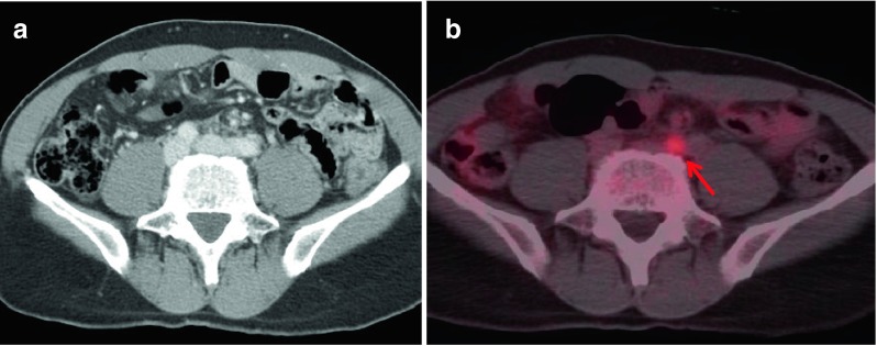
By CeCT (a), there was no definite abnormal finding. However, the PET/CT image (b) showed an additional hypermetabolic lymph node in the left common iliac area (red arrow, SUVmax: 2.2, size: 0.7 cm) (patient no. 6)
Official websites use .gov
A
.gov website belongs to an official
government organization in the United States.
Secure .gov websites use HTTPS
A lock (
) or https:// means you've safely
connected to the .gov website. Share sensitive
information only on official, secure websites.
By CeCT (a), there was no definite abnormal finding. However, the PET/CT image (b) showed an additional hypermetabolic lymph node in the left common iliac area (red arrow, SUVmax: 2.2, size: 0.7 cm) (patient no. 6)