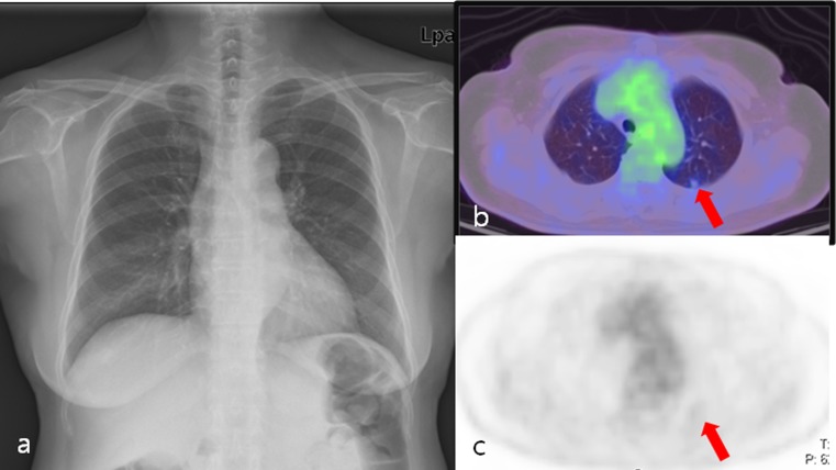
Fig. 2.
A 70-year-old female postoperative rectal cancer patient. a Chest X-ray showed no abnormal findings. b, c Axial PET/CT fusion and PET images showed a solitary nodule (arrow) with faintly perceptible FDG uptake in the left upper lobe (SUVmax: 0.8). Wedge resection confirmed metastatic adenocarcinoma

