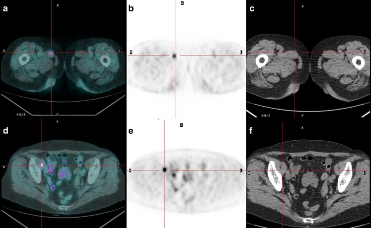
Fig. 1.
18F-FDG PET/CT confirms the presence of the lesion revealed by 131I scan and shows also two muscular distant lesions, involving the right adductor longus muscle (a PET/CT fused transaxial images, b PET transaxial images, c CT transaxial images) and right iliopsoas muscle (d PET/CT fused transaxial images, e PET transaxial images, f CT transaxial images). The lesions were confirmed as metastases from papillary carcinoma by biopsy. To the best of our knowledge, this is also the first described case of a double distant muscle metastasis imaged with 18F-FDG PET/CT

