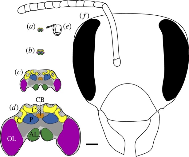
Figure 1.

Stylized cross-sectional schematics of worker brains and heads, illustrating the approximate size range of taxa included in our study. Scale bar, 500 μm for all images. Brains of the ants (a) Brachymyrmex depilis and (b) Tapinoma sessile; (c) brain of the wasp Mischocyttarus mastigophorus; (d) brain of large worker of the bumblebee Bombus impatiens, with brain regions analysed in this study indicated; (e) B. depilis ant worker head; (f) B. impatiens large bumblebee worker head. Key to indicated brain regions (colour references refer to online figure): OL, optic lobe neuropil (purple); AL, antennal lobe neurophil (green); C, mushroom body calyx neuropil (yellow); P, mushroom body peduncle neuropil (blue); CB, central body neuropil (orange); other protocerebral neuropils, dark grey; Kenyon cell body region, stippled light grey. Schematics are based on images from this analysis, published studies [37,49,52] and online sources (discoverlife.org and antweb.org). (Online version in colour.)
