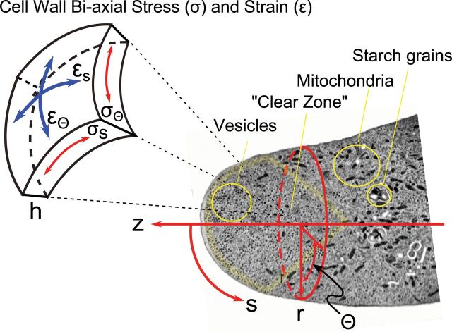
Figure 2.
Coordinate Systems for Pollen Tube Growth Models.
An electron micrograph (Lancelle and Hepler, 1992), labeled to show the location of mitochondria, starch grains, secretory vesicles, and the ‘clear zone’ is overlain with a cylindrical coordinate system in which elongation is measured along the z-axis while the variable r(z) specifies the location of the cell wall along z and thus cell shape. Since the cell is assumed to be axisymmetric around z, cell shape is also fully described by Кs(S), curvature along the meridian (μm–1). Cell wall properties and dynamics are specified in a unit cell in which tensile stress in the wall (σ, MPa) is followed along two orthogonal directions, circumferential (σθ), and meridional (σs), and the corresponding rates of strain (s–1) are termed έr and έs. Wall thickness (h, μm) is a balance between loss of material by viscous flow and secretion of new wall material by vesicle fusion with the cell membrane. The respective values of the two components of biaxial stress at any point on the surface of the cell depend upon internal hydrostatic pressure, wall thickness, local wall curvature, and the degree of flow coupling (Dumais et al., 2004).

