
Figure 3.
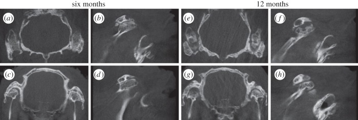
CBCT images of the operated TMJ joints in the six- and 12-month groups represented as (a,e) axial and (c,g) coronal views as well as sagittal views of the (b,f) right and (d,h) left TMJs treated with control and differentiated ASC discs, respectively.