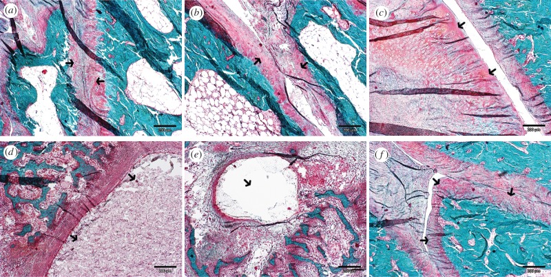
Figure 4.
The sagittal histological sections of the TMJ area stained with Masson's trichrome. (a) The TMJ joint space of the right treated with control ASC disc and (b) the left side treated with differentiated ASC disc in the six-month group. Hyaline cartilage covering the joint surfaces are pointed by arrows. (c) The substantial hypertrophy of condylar cartilage developed in the control ASC disc treated side of the TMJ at 12 months of the follow-up. (d) Microcysts developed in the condylar and (e) temporal bone sides and (f) calcified loose body covered with hyaline-like cartilage are shown in the control ASC disc treated joints after 12 months. The described details are indicated by arrows. Scale bar, 200 µm. (Online version in colour.)

