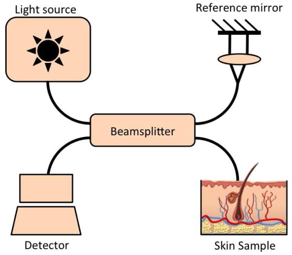
Figure 2.
Schematic of optical coherence tomography system. Light from an optical source is divided by a beamsplitter into 2 fractions. One fraction is guided towards the reference mirror and the other to the tissue. Light reflected from both the reference mirror and tissue are recombined at the beamsplitter and detected to form an interference signal.

