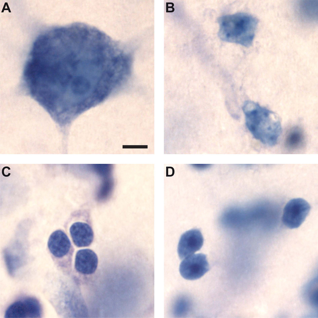
Figure 2.
Classification and identification of different cell types in the basal (A–C) and paralaminar (D) nuclei of the monkey amygdala, viewed with a ×100 objective in Nissl-stained, coronal sections cut at 60 µm. A: Neuron. B: Astrocytes. C: Oligodendrocytes. D: Immature neurons. Scale bar = 5 µm in A (applies to all).

