
Figure 4.
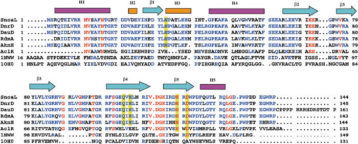
Sequence alignment of SnoaL with other polyketide cyclases and structural homologues, including the secondary structural elements of SnoaL (pink boxes indicate α-helices, yellow boxes 310 helices and arrows denote β-strands). Polyketide cyclases in anthracycline biosynthesis are DnrD (Q54808) from S. peucetius, DauD (Q55215) from Streptomyces sp strain C5, RdmA from S. purpurascens (Q54526) and AknH from S. galilaeus (Q52646). AclR is a protein related in structure, but with unknown function from S. galilaeus (Q8VWA2). A structure-based sequence alignment of the structural homologues limonene-1,2-epoxide hydrolase (1NWW; Arand et al, 2003) and Δ5-3-ketosteroid isomerase (1OHO; Kim et al, 1997) is included. Invariant residues in the polyketide cyclase family are marked in red, and highly conserved residues are shown in blue. Active site residues in polyketide cyclases are highlighted by a yellow background.