
Figure 6.
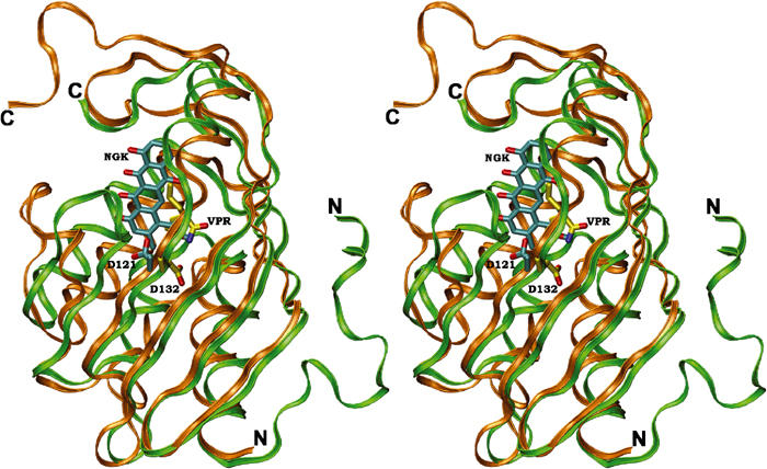
Stereoview of a superposition of the structures of SnoaL (gold) and limonene-1,2-epoxide hydrolase (green). The ligands of SnoaL (NGK) and limonene-1,2-epoxide hydrolase (VPR) are coloured in cyan and yellow, respectively. The position of the structurally conserved aspartic acid residue, involved in proton transfer steps during catalysis in both enzymes, is also shown.