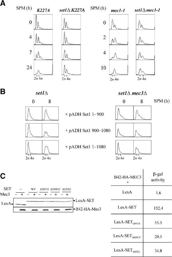
Figure 4.

(A) Meiotic DNA replication is delayed in set1Δ cells independently of a checkpoint response. Meiotic DNA replication in strains with the K227A mutation (left, DBY745 background) and the mec1-1 mutation (right, SK1 background) was followed by FACS analysis. (B) Complementation by the SET domain requires Mec3. set1Δ and mec3Δset1Δ diploids (DBY745 background) were transformed by vectors overexpressing either the full-length Set1 (pADH Set1 1–1080) or fragments of Set1 extending from residue 1 to 900 (pADH Set1 1–900) or from 900 to 1080 (pADH Set1 900–1080) under the control of the ADH promoter (Bryk et al, 2002). Meiotic replication was analyzed by FACS analysis. (C) Interaction of wild-type and mutant SET domains with the C-terminal part of Mec3. Left: expression of the fusion proteins LexA-SET and B42-HA-Mec3 by Western blotting using an antibody against LexA or the HA epitope. Right: protein–protein interaction was monitored by measuring β-galactosidase activity (Miller units). The value represents the average of two measurements. Each measurement was made on five independent clones.
