Fig. 1.
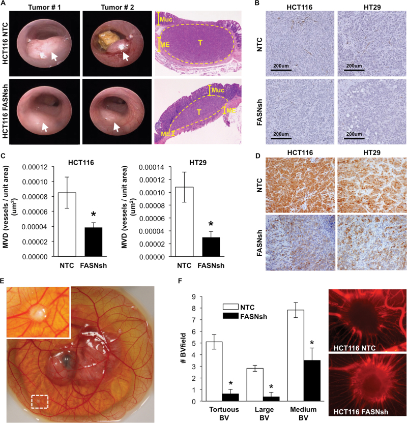
Increased expression of FASN is associated with increased microvessel density in orthotopic colon tumors. (A) Representative images of mouse colon tumors (arrows; 2 weeks) established using HCT116 NTC and FASNsh cells and hematoxylin and eosin staining of tumors and surrounding colon tissues (Muc–mucosa, T–tumor, ME–muscularis externa). (B) IHC staining of CRCs for CD31. (C) MVD analysis using Microvessel analysis algorithm (Aperio ScanScope XT) in eight HCT116 NTC colon tumors (19 sections) and nine HCT116 FASNsh tumors (20 sections), *P < 0.05, and three HT29 NTC colon tumors (nine sections) and three HT29 FASNsh (nine sections), *P < 0.01. (D) Expression of FASN in NTC and FASNsh HCT116 and HT29 orthotopic colon tumors assessed by IHC staining; ×20 magnification. (E) Representative image of the chicken embryo (day 14) with established HCT116 tumors on the CAM. (F) The effect of FASN knockdown on tumor surrounding vasculature in the CAM model. Quantification of tortuous, large (≥25 μm) and medium (10–25 μm) blood vessels surrounding tumors and the representative images of tumors and surrounding vasculature labeled with rhodamine-conjugated lens culinaris agglutinin.

